Introduction
High myopia is characterised by a refractive error of at least -6.00D, or an axial length of over 26.5mm
- Pathological myopia is the excessive axial elongation associated with myopia, which leads to structural changes in the posterior eye that can lead to loss of BCVA.
- Posterior staphyloma is a key feature that contributes to pathological myopia.
- The exact pathogenesis is unclear, but it is proposed that the stretching of the sclera leads to mechanical stress.
Dead Giveaways
Posterior Staphyloma:
Is a deformity or outpouching of posterior globe.
Is a hallmark of pathological myopia, typically leading to a worse prognosis
This predisposes the retina and optic nerve to mechanical damage.
There are 5 main types of staphyloma, with an additional 5 types (6-10) being combinations of the first 5.
Type 1: Wide staphyloma
Type 2: Papillomacular staphyloma
Type 3: Peripapillary staphyloma
Type 4: Nasal
Type 5: Inferior

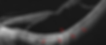
The staphylomas often have such excessive bulging that it shows diffuse choroidal atrophy with visible vessels.

Example of staphyloma

Atrophic Maculopathy:
Progressive chorioretinal degeneration at posterior pole which coalesce to become geographic. This can be thought of as the myopic equivalent of atrophic AMD. Describes the progression of atrophy in 5 separate stages.
A0:
No myopic retinal lesions
A1:
Tessellated fundus only, secondary to thinning of the IP layer, leading to the increased visibility of the choroidal vessels
Low progression chance (15%)

Tessellated Fundus. 
OCT.
A2:
Diffuse chorioretinal atrophy. Can spread to affect entire staphyloma
Yellow-white appearance of posterior fundus
50% progression chance

Chorioretinal atrophy (diffuse) fundus. 
OCT. Shows thin choroid and obvious underlying sclera
A3:
Patchy chorioretinal atrophy
Well-defined white grey areas of atrophy. Loss of RPE and choriocapillaris increases signal transmission
70% progression chance

Myopic maculopathy causing patchy atrophy. 
OCT breakage, as well as increased transmission through to the choroid
A4:
Complete macular atrophy. Complete loss of layers of the macular which can lead to significant loss of vision. Either due to:
Patchy atrophy coalescence and enlargement
CNV related as a secondary cause

Complete macular atrophy. 
Can go through an active phase, scarring phase and finally atrophic.
Tractional Maculopathy:
Traction in myopic maculopathy can be cause retinal, preretinal and subretinal tractions.
Retinal:
ILM rigidity - poor ILM is inability to adapt to changes in elongation.
Choroidal atrophy - poor blood supply to retina and subsequent adhesion between layers.
RPE atrophy
Preretinal:
Anomalous PVD
VMT, ERM
Subretinal:
Traction from staphyloma
T0:
No macular schisis
T1:
Inner or outer foveoschisis
Inner = affecting IPL, GCL, or RNFL
Outer = affecting OPL or ONL

T2:
Inner AND outer foveoschisis

T3:
Foveal detachment (neurosensory detachment from RPE)
T4:
Full thickness macular hole (associated with foveoschisis)

T5:
Macular hole and retinal detachment. IMMEDIATELY REFER

Maculoschisis Grading:

S0 = absent
S1 = extrafoveal
S2 = foveal only
S3 = fovea but not entire macula
S4 = entire macula
Tractional Features:
Vascular Microfolds (20-44.6%)
Retinal vessel inflexibility causes vitreous traction on retina
Inward displacement of tissue around the vessels, thought to be due to the retinal inflexibility

Appears on the surface of OCT, surrounding the vessels
Paravascular retinal cysts (up to 50% of myopic eyes)
Small hollow spaces mainly around large retinal vessels

Appears like cystic spaces
Paravascular lamellar holes (26.8%)
May be due to detachment of the posterior hyaloid or rupture of the paravascular retinal cyst (cyst unroofing)

A form of a pseudohole
ILM detachment (2.4-6%)
Rigid ILM prevents retina from stretching to follow staphyloma

Shows schisis and ILM detachment
Neovascular Maculopathy:
Myopic CNV commonly arise from lacquer cracks (29%), patchy chorioretinal atrophy (20%) and diffuse chorioretinal atrophy (4%), and can reoccur at a different location especially with lacquer crack progression.
N0: [Coin Haemorrhage]
Myopic maculopathy can present without CNV, but still present with haemorrhages.
This is often due to the rupture. Is often associated with lacquer cracks

Liu B et al. 2018. Arrow indicates location of cion haemorrhage. Faintly underneath is the lacquer crack
N1: [Lacquer Cracks]
The lacquer cracks are yellow-white lines at the macula. They represent ruptures in the Bruch's membrane caused by stretching and elongation. This breakage allows vessels to easily enter the choroid
Prevalence of 5-15% in pathological myopia
Associated with CNV or subretinal haemorrhage

Lacquer Crack appearance 
The cracks appear as a hyper-reflective line/column that extends into the choroid and sclera. This may come with a defect in Bruch's membrane
N2a: [Active Phase]
Is characterised by the presence of active growth, identifiable on both OCT-A and OCT

Mixed homogenous and heterogenous reflectivity indicate potential neovascular material. 
From the eye-wiki (AOA). Shows the presence of neovascular and messy vessels in both the avascular zone, and massive proliferative growth in the choriocapillaris. 
A big giveaway is haemorrhaging over the macula. The macula being avascular should not have blood vessels which can cause haemorrhages. This thus reveals myopic maculopathy N2a
N2s: [Scarring Phase]
Characterised by the regression of the blood vessels on OCT, but also the Forster Fuch's spot, which is a pigmented patch.
The consequence leads to atrophic changes, especially A4

Qiuying Chen et al. 2019. Shows the Fuch's spot and the loss of RPE and Bruch's membrane. The large red arrow pointing to the messy heterogenous mix of hyper/hypo reflectivity shows the scarred and regressed CNV. The loss of RPE and Bruch's membrane around it shows the atrophic consequence of CNV (A4)
Dome Shaped Maculopathy:
A convex curved shape elevated profile that is within the concavity of a posterior staphyloma.
Has a prevalence of 10-20%, and is strongly associated with increasing myopia and axial length
Has 3 main types:
Horizontal Oval: Identified on vertical OCT scan, but not horizontal
Vertical Oval: Identified on horizontal OCT scan, but not vertical
Round: Identified on BOTH horizontal OCT scan and vertical

In a horizontally oriented oval, the horizontal line scan will depict a normal retina. But on the vertical OCT, both concave staphyloma and convex dome can be visualised 
Garcia-Ben A, et al. 2017. Displays what the horizontally oriented dome would look like in 3D. 
An opposite effect is seen in the vertically oriented dome, in which case the vertical line scan will depict a normal retina, but on horizontal OCT, both concave staphyloma and convex dome can be visualised. There is also serous detachment. 
Garcia-Ben A, et al. 2017. Displays what the vertically oriented dome would look like in 3D.
diagnostic features
Atrophy Myopic Maculopathy:
The natural history progresses as: Tessellated fundus --> diffuse atrophy --> patchy atrophy --> macular atrophy
Common pattern of progression:
Patchy atrophy enlargement and fusion
Diffuse atrophy enlargement
Diffuse --> patchy atrophy
Tessellated fundus --> diffuse atrophy
Risk of Progression
A1 Tessellated fundus --> 15:85 chance
A2 Diffuse atrophy --> 50:50 chance
A3 Patchy atrophy --> 70:30 chance
Age, female gender, degree of myopia, axial length, presence of posterior staphyloma, lacquer cracks at baseline, worse BCVA baseline.
Tractional Myopic Maculopathy:
The natural history typically is stable. In fact, improvements can usually occur. This is usually due to a complete PVD or spontaneous traction release.
Progression is not associated with age or axial length
Those with severe macular involvement had a higher progression rate. (Some may progress to foveal detachments or FTMH)
Continued monitoring is mandatory.
Neovascular Myopic Maculopathy:
Typically the long term prognosis is very poor, and VA in over 95% can drop to 6/60 within 10 years of CNV onset.
Chorioretinal atrophy is the main cause of the poor prognosis
Furthermore, CNV can develop in fellow eye (35%) within 8 years.
If it presents without CNV (N0), it is typically associated with better prognosis.
Dome Shaped Myopic Maculopathy:
Complications include myopic CNV, serous detachment and macular schisis