Introduction
These lymphoid tumours arise from B and T cell lymphocytes, and represent 6% of all orbital mass lesions.
- Split into 3 categories: A, B and C
- A: Non-specific orbital inflammation (idiopathic orbital inflammation)
- B: Lymphoproliferative reactive
- C: Lymphomas
- 2% of all lymphomas at orbital
- 5-15% of all extranodal lymphomas are orbital
- 50% of all primary orbital malignancies in older adults
- Presents at older ages with a slow onset (50-70s)
- Can be present anywhere in the orbit including the gland
- Has numerous presentations, typically with mucosal-associated lymphoid tissue

Dead Giveaways
Diagnostic Features:
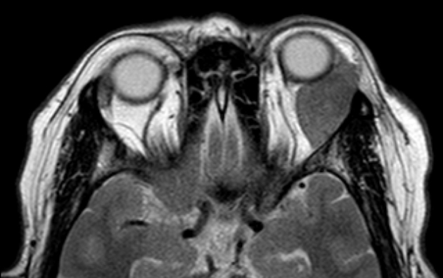
Typically found via imaging techniques such as CT scans or MRI. Can also be diagnosed through histology if a sample can be obtained.
The orbital infiltration is characterised by palpable, firm or rubbery mass
diagnostic features
Symptoms:
Progressive proptosis
Decreased VA
Motility problems
Diplopia
Occasionally will have periorbital oedema